Tomographic Research
The SNRI X-Ray imaging facilities provides academic and industrial users a suite of 3 world-class complementary non-destructive 3-D X-Ray imaging instruments. The SNRI X-Ray Facilities enables researchers from a wide range of disciplines to analyze and characterize material samples as large as 1m in diameter or to image with spacial resolutions of 1mm to 50nm using the highest resolution machines in Anshar.
SNRI Tomographic Anlayses
Reconstruction
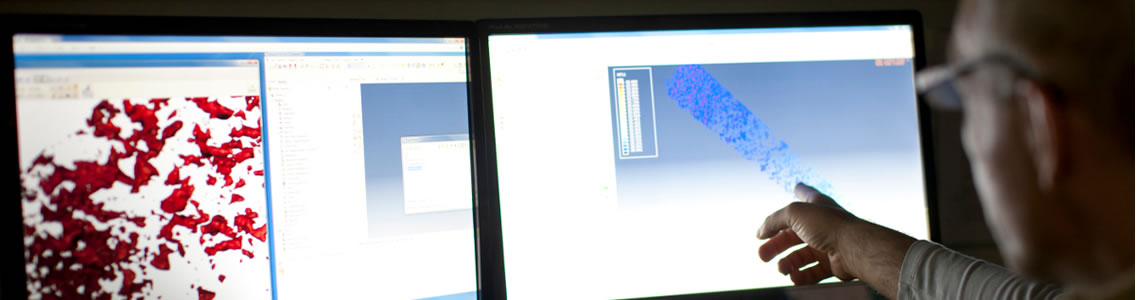
Once your sample has been scanned you will have a large number of radiographs, each of which shows a projection of the 3D object onto the 2D detector. By combining all radiographs it is possible to reconstruct the 3D structure of the object. This reconstruction process also reveals the internal structure of the object. Most of imaging systems have their own custom designed reconstruction software.
For most scientific applications, quantification is key. It is the number, or volume, or distribution of a particular feature within a sample that can answer the important research questions. The SNRI X-Ray group has a range of software packages for 3D visualisation and quantification available for users, some image analysis and quantification tools are available within the major commercial software packages (Avizo, VGStudioMax etc.) and others have been developed to meet a particular need (Dhristi, Quant 3D etc.). The SNRI X-Ray group have a strong emphasis on developing new quantification algorithms in many areas of active research. A member of SNRI staff can advise on your analysis requirements.
New tomographic Method
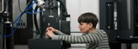
Gamma spectroscopy and gamma emission tomography are two non-destructive measurement techniques for assessing the performance of nuclear fuel which have been investigated in this thesis for existing and novel applications through theoretical studies and experimental demonstrations. For assessment of individual fuel rods using gamma spectroscopy, fuel assemblies are dismantled so that the fuel rods may be measured separately, which is timeconsuming and may cause damage to the fuel.
Key to this research is the Tomographic (X-Ray) Faciliites at SNRI.
Researching Nuclear Fuel
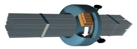
A gamma emission tomography instrument has been designed, constructed and experimentally demonstrated on a Halden Reactor fuel assembly. Simulation studies showed that the instrument and the tomographic reconstruction methods employed may be useful for: identifying a leaking fuel rod in an assembly by its lack of fission gas content; reconstruction of the rod-wise fission product distributions in the fuel stack and plenum regions of the assembly; and determining the rod-wise fission gas release fractions.
Fuel Cycle
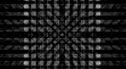
The current use of manual inspection by fuel vendors for quality control of the fabricated fuel pellets is not only-time consuming but also exposes the inspection staff to limited radiation. SNRI is developing an automated, real-time fuel rod inspection technique for better speed and accuracy using an X-Ray tomographic inspection technique to detect anomolies. This higher quality check is enabling SNRI to further develop and research new pathways into fuel recycling.
>> Read Morecontact us
Have questions? Need information? Looking for employment? SNRI is constantly seeking to engage with driven, innovative, and team oriented researchers, analysts, engineers. Contact us about potential research projects and joint opportunities!